
Pregenetic Diagnosis and Screening [PGD & PGS]
Preimplantation Genetic Diagnosis (PGD) was developed in the late 1980s as an alternative to prenatal diagnosis and possible termination of pregnancy of an affected fetus for couples who are at risk of passing on serious genetic diseases to their children.
Preimplantation Genetic Diagnosis technique requires the use of the test tube baby technique (IVF) to test embryos for genetic disorders before it implants in the womb (uterus). It avoids the need for abortion. The procedure is associated with ethical and medical concerns and raises issues of sex selection and genetic engineering.
Despite an increasing number of genetic disorders that can now be diagnosed by this technique, not all-genetic disorders can be diagnosed in this way.
It should be noted that genetic disorders could be due to either a single gene disorder or sex linked chromosomal abnormalities. In single gene disorders, such as cystic fibrosis, the actual genes of the sampled embryo can be examined for the presence of the condition. Other genetic disorders, such as Duchenne’s muscular dystrophy, or hemophilia, affect only males (sex linked diseases). In these cases, the cell is examined to determine the sex of the embryo and only female embryos are replaced.
In cases of recurrent chromosomal abnormalities such as Down’s syndrome and recurrent miscarriages caused by parental translocations, the number and character of several chromosomes can be determined.
How is PGD performed?
Standard IVF technique is used when the procedure requires FISH analysis. ICSI is used when there is abnormal semen or PCR analysis is required. This is to reduce the risk of contamination with sperm DNA.
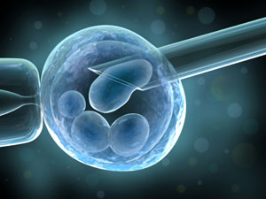
A single cell is removed from an 8-cell embryo through an opening in the outer protective coat. The procedure is carried out under the microscope without damaging its ability to continue to develop normally (because at this stage of development none of the embryo cells have become specialized). The cell is then analyzed for the presence of genetic disorders.
A new technique, called comparative genomic hybridisation (CGH), allows doctors to remove cells from the embryo at a later stage, when it is five days old and has about 100 cells. Removing cells at this stage should be less damaging, and by analysing five or six cells the clinician can be more confident that the genetic abnormality exists in the embryo.
To whom PGD is advised?
- Couples with repeated pregnancy loss due to genetic disorders.
- Couple who have a child with a genetic disease and are at high risk of having another.
- Couples who wish to identify a tissue match for a sick sibling who can be cured with transplanted cells.
Couples with genetic disorders should receive adequate counselling before embarking on PGD. They should be informed about other options such as prenatal diagnosis and termination of pregnancy if the fetus is affected, gamete donation, remain childless and adoption. They should also be counselled about the risks of misdiagnosis and no diagnosis be made.
Unfortunately, for older women and women with high baseline FSH levels, PGD may not be a good alternative because of lower pregnancy rates. Also, they may not produce enough embryos to be tested.
Clinical experience remains limited and the test is not 100% reliable, as sometimes the analyzed cell does not represent the rest of the embryo. To date, about 1000 babies have already been born worldwide using PGD. There are no reports of increased fetal abnormalities following PGD. However, long-term consequences on the fetus is unknown at present.
It is important to note that there is a possibility that none of the embryos tested will be normal, in this case none would be suitable to transfer. Furthermore, as with all tests there is a possibility of a false positive and false negative result (1 in 6 and 1 in 50 respectively). This is because many embryos display inconsistency in chromosomes from cell to cell (mosaicism) and thus the cell taken for biopsy may not be a representative of the other cells of the embryo. For this reason, it is recommended that woman be tested during pregnancy (at 11-16 weeks) for chromosome abnormalities.
. The table below shows examples of genetic diseases that can be diagnosed using PGD.
| Autosomal Recessive | Autosomal Dominant | Sex-linked | Chromosomal Abnormalities |
|---|---|---|---|
| Cystic Fibrosis | Huntingtons Disease | Hemophilia | Translocation |
| Tay Sach’s Disease | Congenital Adrenal Hyperplasia | Duchenne Muscular Dystrophy | Klienfelters Syndrome |
| Thalassemia | Marfans Syndrome | Fragile X | Aneuploidy |
| Sickle Cell Disease | Myotonic Dystrophy | X Linked Mental Retardation | |
| Spinal Muscular Dystrophy | Wiskott-Aldrich syndrome | ||
| Breast cancer (BRCA1) | |||
| Familial Adenomatous polyposis coli (FAP) | |||
| Familial Alzheimer’s disease |
Pre-implantation genetic screening (PGS)
Pre-implantation genetic screening (PGS), also called aneuploidy screening, involves screening embryos using FISH for common aneulopidy (chromosome 21, 18, 16, 13). Aneuploidy is a condition where the embryo contains the wrong number of chromosomes in each cell. The doctor then selects only the chromosomally normal embryos for embryo transfer. In the UK the procedure is licensed for use in older women where the incidence of chromosomal abnormalities such as Down’s syndrome increases by seven to seventy fold, women with a history of recurrent miscarriages, or repeated IVF failure (with good quality embryos) in order to increase the IVF live birth rates. Evidence is lacking that PGS improves the live birth rates in older women and two recent randomised trials found no improvement in live birth rates in older women.